× close
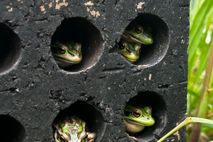
Light microscopy images of histologically stained sections of stem roots. A thin wavy sheet of non-living stem roots surrounded by cilia (red) is seen embedded in the generator’s living tissue (light blue). Credit: Jenaes Sivasundarampillai
A team of chemists from McGill University, in collaboration with colleagues from Germany’s Charité University, have uncovered some of the processes that mussels use to attach to rocks and then quickly detach from them when conditions are right.
In their project, report in diary scienceThe research group studied the interface between the mussel’s tissue and the bundles of filaments that the mussel uses to anchor itself to rocks and other objects. Guoqing Pan and Bin Li from Jiangsu University and Dongzhou University in China. perspective article The same journal issue provides an overview of the work the team has done on this new initiative.
Mussels are bivalve molluscs that live in both freshwater and saltwater environments. They have a hinged shell connected by ligaments. Once the shell is closed, it is tightly sealed by muscles. Mussels use byssus threads (commonly known as barbels) to attach themselves to solid objects, such as rocks.
Mussel byssus has been extensively studied due to its unique ability to connect and optionally sever abiotic materials (the filaments that make up the thread) to biological tissue. However, as Pan and Li point out, most of this research revolves around possible chemical bonding mechanisms. In this new effort, the research team focused instead on the dynamics of the biointerface.
To better understand how the byssus connects to living tissue and, if necessary, how it is disposed of, the research team used a variety of techniques to isolate the byssus and the tissues it connects to. I researched. Using a combination of imaging and spectroscopy techniques, the researchers observed that the ends of the threads were intertwined with layers of biological tissue and were themselves covered with about 6 billion motile cilia.
× close
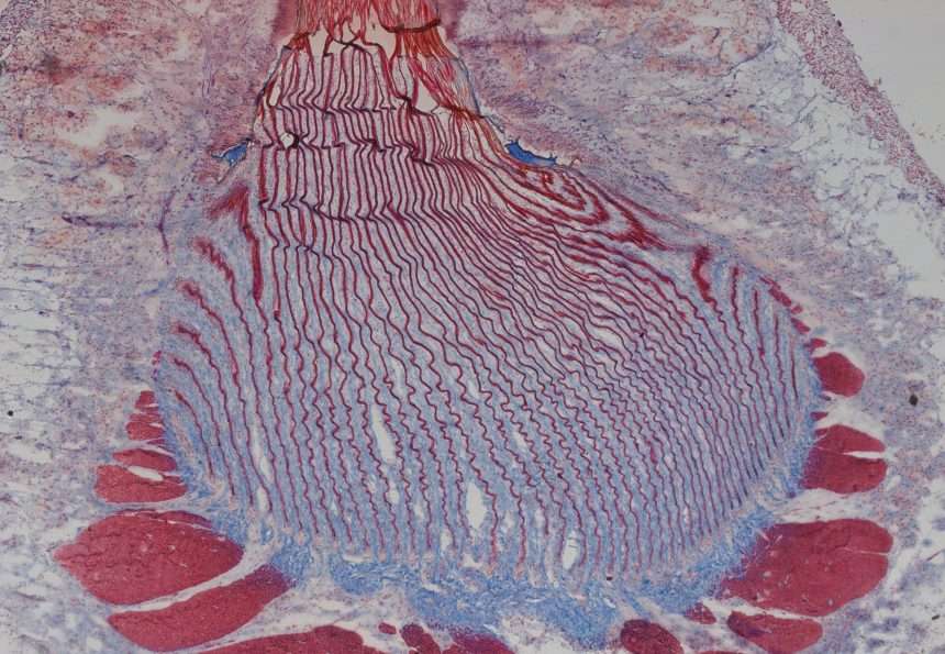
Features reconstructed in 3D from a FIB-SEM image stack created from a small area at the base of the stem. Living tissues are dark blue, non-living stem root sheets are light blue, secretory vesicles are blue-green, and cilia are red. Credit: Jenaes Sivasundarampillai
They further discovered that the large number of cilia provides a high degree of surface contact, which makes it possible to mechanically interlock two different materials. The researchers also noted that the cilia’s vibrations helped strengthen the grip between the two materials, allowing for rapid release when needed. Researchers have discovered that cilia movement is driven by neurotransmitters, and researchers theorize that cilia are ultimately controlled by serotonin and dopamine.
For more information:
Jenaes Sivasundaramillai et al. A strong quick-release biointerface in mussels mediated by serotonergic cilia-based adhesion, science (2023). DOI: 10.1126/science.adi7401
Guoqing Pan et al. Dynamic biointerface controls mussel adhesion; science (2023). DOI: 10.1126/science.adl2002
© 2023 Science X Network