Sign up for CNN’s Wonder Theory science newsletter. Explore space with news of fascinating discoveries, scientific breakthroughs, and more.
CNN
—
An extinct marine creature, ribbon-like and about the size of a human hand, was one of the earliest animals to evolve a precursor to a spine, and scientists recently identified the animal’s nerve cord by twisting it upside down: they turned the fossil upside down.
Paleontologist Charles Doolittle Walcott Pikaia fossils were first found in the Burgess Shale of British Columbia, dated to 508 million years ago, and described in a 1911 paper. The animal was about 6.3 inches (16 cm) long, with a flattened, sinuous body and a small head terminating in two tentacles fringed with external gills. These were originally thought to have been primitive legs, and the animal was positioned so that these structures pointed downwards.
In 2012, after decades of studying the Pikaia fossils, researchers Explained The researchers examined the fossil’s internal structure in great detail, identifying long threads near the abdomen as blood vessels and a sausage-shaped structure running down the animal’s back that they named the dorsal organ. The dorsal organ was probably used for internal support, but such an organ was anatomically different from anything seen in fossils or living animals.
But a more recent analysis of Pikaia fossils by a different team of scientists was published in the journal Nature on June 11. Current Biologyoverturned this view and all previous research on Pikaia.
Previous anatomical interpretations had positioned the animal inside-out, the researchers say: the so-called dorsal organ was actually in the abdomen and Pikaia’s intestine, and what were thought to be blood vessels were actually nerve cords, characteristic of a group of animals in the phylum Chordata known as chordates.
Giovanni Mussini
An annotated photograph shows the newly revised configuration of Pikaia gracilens. The abbreviations in Box C indicate the main features of the fossils seen in Box B: the head tentacles of Pikaia (Tc), dorsal nerve cord (In), dorsal nerve cord (Nc), possible gonads (?Go), and the myoseptum, or connective fascia (Ms). The diagram in Box G indicates the features of the fossils in Box F: the forelimbs (Aa), pharyngeal cavity (Ph), intestinal canal (Gu), and myotome, or muscle segment (My). The fossil specimens are from the Smithsonian National Museum of Natural History, except for the fossils in Box I, which are from the Royal Ontario Museum.
Vertebrates, chordates such as eel-like amphioxus and sea squirts, all develop a flexible, rod-like nerve structure called a notochord on their backs at some point in their lives.
Pikaia was originally thought to be a worm, but was later classified as an early type of chordate based on characteristics such as certain muscle shapes and the position of the anus, but experts weren’t sure exactly where Pikaia fit on the chordate family tree.
The description of the nerve cord means that Pikaia is now considered to be part of the basal lineage of all chordates, even though it has no living direct descendants, the study authors reported.
The Pikaia paradox is “very revealing,” say evolutionary biologists. Dr. John MallattMallatt, a clinical professor at the University of Idaho, was not involved in the new study but has published papers on Pikaia. In 2013working from an established (and inverted) body position.
In retrospect, the truth was “hiding in plain sight,” Mallatt said, and this change of direction clears up the question of why Pikaia’s vascular and dorsal structures clashed with well-established anatomical features of other chordates.
“Suddenly Pikaia doesn’t seem so strange,” he said.
The reevaluation of which is correct for Pikaia began several years ago, with the co-authors of the new study. Dr. Jacob Winter“These findings suggest that the evolution of primates is a fundamental step forward for the evolution of mammals,” said lead study author David L. Schneider, a lecturer in macroevolution at the University of Bristol in the UK. Giovanni Mussiniis a researcher and PhD student in the Department of Geosciences at the University of Cambridge, UK.
Mussini told CNN there are several reasons to reconsider previous interpretations of the fossil, one of which is a mystery about what scientists believed to be the location of a dorsal organ, which is near what is believed to be Pikaia’s back, seemingly ruling out the possibility that the organ was an intestine.
However, when Pikaia was turned over, the location and features of its organs made more sense anatomically. The organs had widened and extended into the animal’s pharynx, the part of the throat where the intestine joins the mouth. Their three-dimensional state can be explained by the presence of highly chemically reactive tissue, characteristic of the intestine. In other Burgess Shale fossils, the abundance of ions and reactive compounds typically found in intestinal tissue caused the digestive organs to mineralize faster than the rest of the body, and therefore retain more of their original shape. Research has shown that the internal structures of Pikaia’s organs may have been remnants of swallowed food.
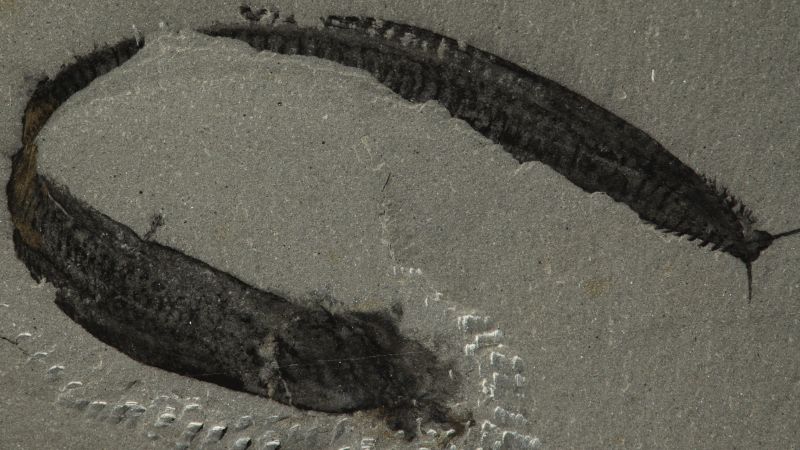
Giovanni Mussini
This image of a Pikaia fossil specimen at the Smithsonian National Museum of Natural History shows the intestine, masses of muscle tissue called myotomes, and a dorsal nerve cord, with light-colored deposits visible inside the intestine (toward the head on the right).
In the upside-down Pikaia, the external gills, which previously faced downwards, are now similar to those of modern mudskippers and Axolotl.
Pikaia’s flip also reoriented the undulating muscle groups, called myosea, which are a key feature of vertebrates. In Pikaia’s new position, the strongest bending points of these muscles are along its back, which is consistent with the arrangement of myosea in other vertebrate animals.
“Pikaia’s movements are consistent with those seen in modern chordates,” Mussini said.
Pikaia’s putative blood vessels were also anatomically puzzling, lacking the branching structures typical of vertebrate blood vessels.
“It’s a single line that runs through most of the body up to the head, where it branches into two threads that become tentacles,” Mussini said.
Giovanni Mussini
An illustration of the head of Pikaia gracilens from a fossil specimen at the Smithsonian National Museum of Natural History highlights the thickening of the dorsal nerve cord. The discovery of other fossil nervous systems from the Cambrian period has allowed scientists to reassess how Pikaia was organized.
A key part in realizing that this structure was a nerve cord was the fossil nervous systems of other Cambrian animals (541 million years ago to 485.4 million years agoMussini went on to mention some of the many celestial objects that have been discovered in the past decade.
“We’ve been fortunate to find quite a few Cambrian nervous systems preserved in other deposits, which has improved our understanding of how nerve cords and other tissues fossilize,” he said. “Most of these are Chinese fossils that have been found in the last few years.”
Many of these fossils were arthropods (invertebrates with an exoskeleton), which include modern-day relatives such as insects, arachnids, and crustaceans. By comparing the fossils to modern arthropods, paleontologists were able to identify preserved internal tissues. One example is a fossil specimen of the Cambrian arthropod Morrisonia, which showed brain tissue comparable to that of modern spiders, scorpions, and horseshoe crabs, Mussini said.
Although there are no organisms similar to Pikaia, the fossil arthropod data have given scientists a more detailed frame of reference for Pikaia’s nerve cord. Like other fossilized nervous tissues, Pikaia’s nerve cord was dark, high in carbon, and relatively fragile compared to other fossilized tissues.
This nerve cord firmly establishes Pikaia’s status as a chordate, “putting it almost at the base of what we think of as traditional chordates,” Mallatt said.
Much about Pikaia’s anatomy remains a mystery, but looking at it from a new angle could provide new insight into its array of puzzling features, Mussini said.
“Many of these details have only emerged in the last 10 or 12 years,” Mussini added, “and the authors of the 2012 paper can certainly be forgiven for not bringing these details into the discussion, because this is ongoing research.”
Mindy Weisberger is a science writer and media producer whose work has appeared in Live Science, Scientific American, and How It Works magazine.