summary: Researchers have identified microfold cells, or M cells, normally found in the intestine, in the thymus gland, an organ essential for the development of the immune system. These thymic M cells resemble their intestinal counterparts and may play a role in immune responses throughout the body. This discovery provides new insights into the complex interactions and functions of organs.
Important facts:
- M cells are primarily known to reside in the intestine, but were unexpectedly discovered in the thymus by Israeli researchers and verified by Professors David Lo and Diana del Castillo.
- Thymic M cells are similar to M cells found in the intestine and airways, but have a different developmental origin.
- The presence of these M cells in the thymus provides valuable insight into the organ’s role in shaping immune responses, similar to processes seen in other parts of the body.
sauce: UCR
Professor David Roe and graduate student Diana del Castillo were recently approached by Israeli researchers for their expertise on specialized cells called microfold cells (M cells), which are primarily known to reside in the intestinal epithelium. When he received the consultation, he was surprised by this. .
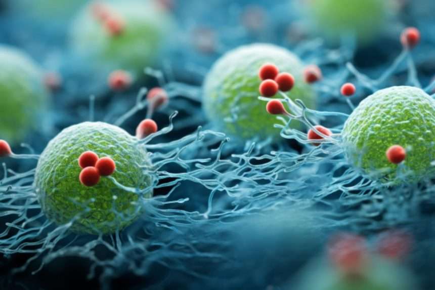
A research group in Israel has identified similar cells in the thymus. The thymus is an organ just above the heart that plays a key role in the immune system and produces lymphocytes, white blood cells that protect the body from infection.

Lo and del Castillo are distinguished professors of biomedical sciences at the University of California, Riverside School of Medicine. Nature, it was confirmed that newly discovered cells in the thymus gland look exactly like M cells. Her M cells, which act like gatekeepers, are specialized antigen-delivery cells for the immune system in organs such as the intestines and lungs. They play an important role during the development of the body’s immune system.
Researchers at Israel’s Weizmann Institute of Science, led by Jakub Abramson, began studying the thymic epithelium in mice before contacting Lowe. Low’s research interests include understanding how M cells in the gut and airways work to build the immune system.
“I have been working on these cells for several years, so when I was contacted by the Israeli team, I was intrigued,” Low said.
“This group is conducting research into the cellular structure of stromal cells (cells that make up certain types of connective tissue) within the thymus, and using new and advanced methods, it is possible to We learned that we had discovered a very similar population of cells” found in the intestines and respiratory tract. In my own research, I never thought of looking for her M cells in the thymus. ”
Fortunately for the Israeli scientists, del Castillo, under Lo’s guidance, studies mouse mucosal tissue (the tissue that lines some body tubes and organs) in the lab. We were able to answer several questions, such as where in the thymus gland is located. Where are the newly discovered cells located and what are they doing there?
“These specific M cells are restricted to specific regions of the thymus and have unique relationships with different cell types and functions,” del Castillo said.
“Questions these cells are already raising include how similar they are to M cells elsewhere in the body, and what makes them different where they are found. ”
Lowe explained that the thymus has long been a tissue of interest to immunologists because most of the development of the immune system is centered on and dependent on the thymus.
“This remains a deep, ongoing mystery that continues to generate interest,” he said. “The thymus provides clues to how the immune system begins. This complex organ has so many different stromal cell types and interactions that protect us from infection. Responsible for the production of protective lymphocytes.
Low said the newly discovered M cells are very similar to those found in the intestines and airways.
“However, thymic M cells have a distinct developmental origin, which is an interesting puzzle in itself,” he says.
“Once they develop, they look very similar to what we’ve been studying in the gut. As you know, M cells capture viruses and bugs that have entered the respiratory tract and pass them to the immune system, where they can be used by the immune system. The system responds to infectious agents. Are M cells doing the same thing in the thymus in terms of organization and function? That’s what we want to know.”
Del Castillo, who is pursuing a doctorate in biomedical science, used genetically modified mice to answer questions from Israeli researchers.
“We found that new cells were scattered in the medullary region of the thymus,” she said. “This has interesting implications in terms of the role and compartmentalization of the thymus, including how these cells function to regulate lymphocyte training within this organ.”
Low and del Castillo were surprised to discover that many of the steps involved in forming immune responses in different parts of the body appear to be mirrored in the thymus.
“It is interesting to see that many of these early cellular interactions and developments that we have studied closely in the peripheral immune system are occurring in the thymus,” Professor Low said.
“We didn’t expect to see these interactions here. It’s like watching a short video in the thymus about what’s happening on a large scale in the periphery.”
The thymus also prevents lymphocytes from accidentally attacking our own tissues. The thymus medulla is where these decisions are made, UCR scientists said.
“The newly discovered M cells are part of this decision-making process,” del Castillo said.
“The production of antibodies in the peripheral immune system to fight infectious microorganisms involves several steps, and many cells interact with each other. What is interesting is that some of these interactions occur in the thymic M This is something that is reproduced in the early stages of cell development.”
Lowe said thymic M cells are thought to be later trained to function in the periphery as needed, allowing them to communicate and interact with other cells.
“The thymus is complex because it produces an entire functional immune system and repertoire, and we know that many components play a role in its performance,” he said. “We did not expect M cells to appear in the thymus. Therefore, this is clearly linked to similar processes occurring in the intestines and respiratory tract, where 60-70% of infectious agents enter the body. is a gratifying discovery.”
About this neuroscience research news
author: Iqbal Pittalwala
sauce: UCR
contact: Iqbal Pitalwala – UCR
image: Image credited to Neuroscience News
Original research: Closed access.
“Thymimimetic cells function beyond self-toleranceWritten by David Roe et al. Nature
abstract
Thymimimetic cells function beyond self-tolerance
The development of immunocompetent T cells in the thymus is necessary for effective defense against all types of pathogens, including viruses, bacteria, and fungi. To achieve this objective, T cells undergo a very rigorous educational program in the thymus, in which both non-functional and self-reactive T cell clones are eliminated by positive and negative selection. .
Thymic epithelial cells (TECs) play an essential role in these processes, and previous studies have shown remarkable heterogeneity of these cells.
Here, we use multi-ohm analysis to provide further insight into the functional and developmental diversity of TECs in mice, revealing a detailed atlas of TEC compartments according to the transcriptional state and chromatin landscape of the cell. Our analysis highlights unconventional TEC subsets that resemble functionally well-defined parenchymal populations, including endocrine cells, microfolded cells, and myocytes.
By focusing on endocrine and microfold TEC populations, we show that endocrine TECs are required. Instagram 1 important for their development and for maintaining thymic cellularity in a ghrelin-dependent manner. In contrast, microfold TEC requires: spiv required for their development and essential for the production of thymic IgA+ Plasma cells.
Taken together, our studies demonstrate that medullary TECs have the potential to differentiate into different molecularly distinct and functionally defined cell types, and that these cells may be involved in the induction of central tolerance. In addition to contributing to the homeostasis of other populations present in the thymus, we have also shown that it regulates the homeostasis of other populations present in the thymus.