
artistic reconstruction Kirinshia. Credit: X. Wang
Researchers from the University of Leicester and the Yunnan Key Research Institute of Paleontology have used advanced scanning technology to recreate a “fossil monster” that lived 500 million years ago.
A joint team including researchers from the University of Leicester, Yunnan Key Research Institute of Paleontology, Yunnan University Institute of Paleontology, Chengjiang Fossil Museum, and Natural History Museum, London, discovered a unique fossil animal discovered about 520 million years ago. was re-examined. old rock. This reassessment helps fill knowledge gaps in the evolutionary history of animals known as arthropods.
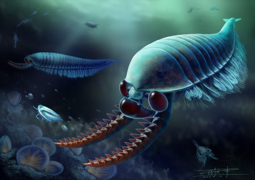
animals with scientific names Kirinshia, Imaging using a CT scanner revealed soft anatomical structures buried in the rock. About the size of a large shrimp, its amazing features include her three eyes on her head and a pair of fearsome limbs, presumably used for catching prey.
This study was recently published in the journal current biology.

CT images of fossil animals Kirinthia jangi Originally from southern China. This animal is about the size of a large shrimp, with its front end on the right side. This image clearly shows the division of the body and the large eyes in the front.Credit: Provided by Professor Yu Liu of Yunnan University
Fossils of various types of marine animals first appeared in rocks about 500 million years ago, marking a time when complex ecosystems were developing in the world’s oceans. One important source of such fossils is the area around the city of Chengjiang in southern China, where the fossils were collected by the Chinese team in this study. The fossils were recovered from the Cambrian Chengjiang biota of Yunnan Province, China. seed A number of exceptionally preserved fossil organisms are described.
This new discovery is important in elucidating the history of arthropods. These are animals whose bodies are divided into several parts, like crabs, lobsters, insects, and spiders, and most of them have a pair of jointed limbs.

CT images of fossil animals Kirinthia jangi Originally from southern China. This image shows the large forelimbs extended.Credit: Provided by Professor Yu Liu of Yunnan University
Although many arthropods (most famously trilobites) are present in the fossil record, most preserve only their hard skeletons. The new Chinese material is almost completely preserved, so the team was able to image the head. KirinshiaSix body segments are identified. The front segment has eyes, the second segment has a pair of large grasping limbs, and each of her other four segments has a pair of articulated limbs.
Dr Robert O’Flynn, lead author of the study and student in the School of Geography, Geology and Environment at the University of Leicester, said: “The preservation of this fossil animal is amazing. After the CT scan, we can digitally turn around and literally look into the face of something that lived over 500 million years ago. Like many extant arthropods, we found that it had six segments on its head.”

Micro CT model Kirinshia It represents the features of the head.Credit: Courtesy of YKLP Professor Yu Liu
Professor Mark Williams, Robert’s main supervisor at the University of Leicester, said:Kirinshia, and the Sumjiang biota from which it originated, will help build our understanding of early euarthropod evolution. I believe similar discoveries will be made by Robert in the future. ”
Professor Yu Liu of the Yunnan Key Research Institute of Paleontology said: “Robert and I had been examining micro-CT data as part of our doctoral thesis in hopes of refining and correcting previous interpretations of the head structure of this genus Chirinthia. It turns out that it is made up of six parts, just like insects.
Dr Greg Edgecombe from the Natural History Museum added: “Most of our theories about how arthropod heads evolved are based on these early divergent species, which had fewer body segments than extant species. Previously undiscovered 2 found a pair of legs Kirinshia The findings suggest that modern arthropods inherited six-part heads from their ancestors at least 518 million years ago. ”
Reference: “Early Cambrian Qilinthia Zangi and the Evolution of Arthropod Heads” Robert J. O’Flynn, Yu Liu, Xianguan Hou, Huijuan Mai, Mengxiao Yu, Songlin Chuan, Mark Williams , Jing Guo and Gregory D. Edgecombe, 28 August 2023 current biology.
DOI: 10.1016/j.cub.2023.08.022
This research was funded by the Yunnan Science Foundation.